Some consider it to be part of the brainstem though most treat it as a portion of the forebrain. The diencephalon includes the dorsal thalamus, hypothalamus, ventral thalamus, and the epithalamus, and it is situated between the telencephalon and the brainstem. In general, the diencephalon is the main processing center for information destined to reach the cerebral cortex from all ascending sensory pathways (except those related to olfaction) and numerous other subcortical cell groups. The right and left halves of the diencephalon, for the most part, contain symmetrically distributed cell groups separated by the space of the third ventricle (Haines 190).
The dorsal thalamus is the largest of the four principal subdivisions of the diencephalon and consists of pools of neurons that collectively project to all areas of the cerebral cortex. Some of the thalamic nuclei receive somatosensory, visual, or auditory input and transmit this information to the appropriate are of the cerebral cortex. Other thalamic nuclei receive input from subcotical motor areas and project to those parts of the overlying cortex that influence the successful execution of a motor act. A few thalamic nuclei receive a more diffuse input and, in return, relate in a more diffuse way to wide spread areas of cortex (Haines 190)
Fan-shaped mass of fibers of axons that pass between the diencephalon, particularly the dorsal thalamus, and the cerebral cortex. Passes from the central core of the hemisphere in the brainstem (Haines 197). In an axial plane through the hemisphere, the internal capsule appears as a prominent V-shaped structure. It is divided into three parts:
Anterior limb-Insinuated between the head of the caudate nucleus and the lenticular nucleus.
Posterior limb-Located between the dorsal thalamus and the lenticular nucleus.
Genu-Located at the inter-section of the anterior and posterior limbs, which is approximately at the level of the interventricular foramen (Haines 210-211).
nuclei that project to a single, functionally uniform region of cortex from VL, VPL, VPM and geniculate nuclei of the thalamus.
somatosensory info is received here in this section of the thalamus. The medial lemniscus and spinothalamic fibers terminate in a somatotropic manner in the VPL. The VPL then projects axons to the postcentral gyrus of the frontal lobe.
The trigeminal tract (sensory info from the face) and the spinal trigeminal nucleus terminate in the VPM which then synapses and sends axons to the postcentral gyrus of the frontal lobe.
Composed of multiple nuclear subdivisions and is connected primarily to the forebrain, brainstem, and spinal cord. This part of the diencephalon is involved in the control of visceromotor (autonomic) functions. In this respect, the hypothalamus regulates functions that are "automatically" adjusted (such as blood pressure, temperature, etc.) without our being aware of the change. In contrast, conscious sensation and voluntary motor control are mediated by the dorsal thalamus (Haines 190).
X-shaped. Carries optic nerves from the retina. At the optic chiasm, fibers from the medial aspect of each eye crossover to the opposite side and then continue on via the optic tracts (Marieb 560). On a midsagittal view, the optic chiasm can be seen at the ventral-rostral point of the hypothalamus.
Between the optic chiasma and mammillary bodies os the infundibulum, a stalk of the hypothalamic tissue that connects the pituitary gland to the base of the hypothalamus (Marieb 421). The pituitary gland produces 9 major hormones including Oxytocin, ADH, TSH, and GH.
"Arch". Fiber tract that link the limbic system regions together (Marieb 428).
Paired pea-like nuclei that bulge anteriorly from the hypothalamus, are relay stations in the olfactory pathways (Marieb 421). The nuclei receive input via the fornix. The mammillary nuclei are involved in the control of various reflexes associated with feeding, as well as mechanisms relating to memory formation (Haines 199).
A diffuse group of fibers that courses rostrocaudally through the lateral hypothalamic area. This bundle is complex, in that it conveys ascending inputs into the hypothalamus and through this area into the septal region. It also is a major conduit through which the septal nuclei and portions of the hypothalamus communicate with the brainstem. The dopamine-containing fibers in this area are thought to be related to perceptions of pleasure or drive reduction (Haines 452-453).
Found deep within the cerebral white matter of each hemisphere. Made up mainly of the caudate nucleus, putamen and globus pallidus (Marieb 418).
The smaller medial portion of the lenticular nucleus. It is located internal to the putamen and is smaller in all dimensions. It is divided into medial (internal) and lateral (external) parts by thin sheets of dorsoventrally oriented white matter (Haines 215).
Caudate Nucleus and lenticular nuclei form the corpus striatum. The caudate nucleus is characteristically located in the lateral wall of the lateral ventricle and consists of three parts, a head, body, and tail. The head of the caudate nucleus forms prominent bulge in the anterior horn of the lateral ventricle. At about the level of the interventricular foramen, the caudate diminishes in size, but continues caudally as the body of the caudate nucleus in the lateral wall of the body of the lateral ventricle. In the lateral wall of the collatera trigone, the body of the caudate nucleus turns ventrally then anteriorly to continue rostrally as the tail of the caudate nucleus in the lateral wall of the temporal horn. This part of the caudate is located in the dorsolateral wall of the temporal horn. Thus, the C-shaped of the caudate nucleus faithfully follows the C-shape of the lateral ventricle (excluding the posterior horn) (Haines 213-215).
Part of the lenticular nucleus which is located in the base of the hemisphere. It is the larger lateral part. The smaller medial portion is the globus pallidus. The putamen extends more dorsal, more anterior, and more caudal than does the globus pallidus and is clearly the larger part of the lenticular nucleus when viewed in axial or coronal planes (Haines 215).
Group of structures located on the medial aspect of each cerebral hemisphere and diencephalon. Its cerebral structures encircle (limbus ="ring") the upper part of the brain stem and include parts of the rhinencephalon and part of the amygdala. In the diencephalon, the main limbic structures are the hypothalamus and the anterior nucleus of the thalamus. The fornix and other fiber tracts link these limbic system regions together. The limbic system is our emotional, or affective (feelings) brain. Two parts seem especially important in emotions—the amygdala and the anterior part of the cingulate gyrus (Marieb 428).
Part of the limbic system. It is important in emotions (Marieb 428). The dorsal part of the limbic system.
Through a variety of pathways the Hippocampal formation and amygdaloid complex interconnect with numerous telencephalic and diencephalic centers (Haines 216). Composed of the subiculum, hippocampus, and the dentate gyrus. The subiculum is laterally continuous with the cortex of the parahippocampal gyrus. Medially the edge of the hippocampal formation is formed by the dentate gyrus and the fimbria of the hippocampus (Haines 446). Plays a role in converting new information into long-term memory.
Almond shaped. Amygdaloid nucleus. Sits on the tail of the caudate nucleus. Functionally belongs to the limbic system (Marieb 419).
Part of the rhinencephalon (Marieb 428). A small area just rostral to the anterior commissure and in the medial wall of the hemisphere (Haines 452).
Form the most superior part of the brain. Nearly the entire surface of the cerebral hemispheres is marked by elevated ridges of tissue called gyri, literally "twisters", separated by shallow grooves called sulci, "furrows". It is separated into the frontal, parietal, occipital, temporal lobes and the insula (Marieb 409-410).
Interconnect different areas of cortex within the same hemisphere. They connect the cortices of adjacent gyri (Haines 210).
Interconnect different areas of cortex within the same hemisphere. They interconnect distant areas of the cortex. Examples would be the inferior longitudinal fasciculus (temporal-occipital interconnections), and the uncinate fasciculus (frontal-temporal interconnections). The superior longitudinal fasciculus, located in the core of the hemisphere, interconnects frontal, parietal, and occipital cortices, whereas the arcuate fasciculus interconnects frontal and temporal lobes (Haines 210).
"Radiating Crown". The distinctive arrangement of projection tract fibers from the internal capsule that radiate fanlike through the cerebral white matter to the cortex (Marieb 418).
Commissural fibers that interconnect corresponding structures on either side of the neuroaxis. The largest bundle of commissural fibers is the corpus callosum. Located dorsal to the diencephalon and forms the roof of much of the lateral ventricles (Haines 210).
At the upper limit of the brain stem, the projection of fibers on each side from a compact band called the internal capsule which passes between the thalamus and some of the basal nuclei. Beyond that point, they radiate fanlike through the cerebral white matter to the cortex (Marieb 418).
The rostral most point of this lobe is the frontal pole of the brain. Separated from the parietal lobe by the central sulcus (Haines 207).
On the anterior side of the Central sulcus. It lies in the frontal lobe. Together with the anterior paracentral lobule of the frontal lobe, forms the primary motor cortex (Haines 207).
Lies in the frontal plain. Separates the frontal lobe from the parietal lobe (Marieb 410, Haines 206).
Consists of the postcentral gyrus and the superior and inferior parietal lobules. The postcentral gyrus and posterior paracentral lobule constitute the primary somatosensory cortex (Haines 208).
Gyrus on the posterior side of the central sulcus. It is on the parietal lobe (Marieb 410). It is between the Central and Post-central sulci. Part of the primary somatosensory cortex (Haines 207-208).
Gyri forming the superior parietal lobule extend onto the medial surface of the hemisphere as the precuneus, whereas the inferior lobule is made up of the angular and supramarginal gyri. The latter is a crescent-shaped ridge of cortex around the caudal terminus of the lateral sulcus (Haines 207).
Forms the caudal end of the hemisphere; its caudal extreme is the occipital pole of the brain. The irregular collection of gyri on the lateral surface of the occipital lobe form the occipital gyri. The primary visual cortex is located here (Haines 209).
On the occipital lobe. Separates the cuneus, which is dorsal to it from the lingual gyrus, which is ventrally located. The primary visual cortex is located in the portion s of these gyri that border directly on the calcarine sulcus (Haines 209).
Found on lateral and ventral aspects of the hemisphere between the lateral sulcus and the collateral sulcus. On the upper edge of the temporal lobe, and extending into the depths of the lateral fissure, are the transverse temporal gyri. These form the primary auditory cortex (Haines 208).
long fissure that runs horizontal and separates the frontal and temporal lobes
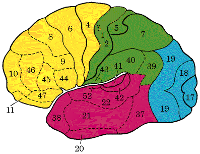
Primary Somatosensory Cortex. Resides in the post central gyrus of the parietal lobe, the anterior most part of the parietal lobe. Neurons in this gyrus receive information, relayed from the general (somatic) sensory receptors located in the skin and from proprioceptors in skeletal muscles. (Marieb 412)
Primary Motor Area. Located in the precentral gyrus of the frontal lobe of each hemisphere. Large neurons, called pyramidal cells, in these gyri allow us to consciously control the movements of our skeletal muscles (Marieb 412).
http://www.umich.edu/~cogneuro/jpg/Brodmann.html
Premotor Cortex. Located just anterior to the precentral gyrus in the frontal lobe. This region controls learned motor skills of a repetitious or patterned nature such as playing a musical instrument and tying (Marieb 413).
Primary Visual Cortex. Seen on the extreme posterior tip of the occipital lobe. Most of it, however, is located on the medial aspect of the occipital lobe, buried within the deep calcarine sulcus. The largest of all cortical sensory areas, the primary visual cortex receives visual information that originates on the retinas of the eyes (Marieb 414).
Visual Association Area. Surrounds the primary visual area and covers much of the occipital lobe. Communicating with the primary visual area, the visual association area interprets visual stimuli in light of past visual experiences, enabling us to recognize a flower or a person’s face and to appreciate what we are seeing (Marieb 414).
Primary Auditory Area. Located in the superior margin of the temporal lobe abutting the lateral sulcus. Sound energy exciting the hearing receptors of the inner ear causes impulses to be transmitted to the primary auditory cortex, where they are related to pitch, rhythm, and loudness (Marieb 415).
